


3i Lattice LightSheet Microscope
Ultra-thin light sheet microscope system with spatiotemporal super-resolution and single-molecule sensitivity for live cell research
➸Overview
3i Lattice LightSheet Microscope
A sweet spot of spatial and temporal resolution
|
Invented by Nobel Laureate Dr. Eric Betzig of the Howard hughes Medical Institute Janelia Research Campus, this microscope has been applied to biological systems spanning four orders of magnitude in space and time. An extremely sensitive primary objective coupled with a custom-designed illumination system allows optical sectioning using extremely low light doses for imaging with unprecedented duration. 3D experiment previously limited by phototoxicity can now be continuously monitored for hours or days. The combination of high spatiotemporal resolution, speed and sensitivity makes the lattice LightSheet the ultimate tool in a new era for living cell microscopy. |
|
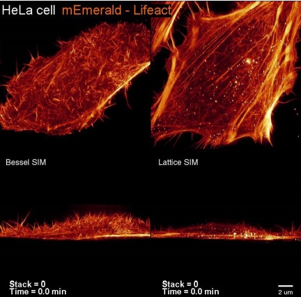
Top and side view volume renderings of HeLa cells expressing mEmerald-Lifeact, demonstrating the higher speed and reduced phototoxicity of the lattice light (right) compared to Bessel beam plane illumination (left) when each is applied to super-resolution structured illumination microscopy (SR-SIM). |
|
|
|
|||
|
|
|||
|
|
|
➸ Advantages
3i Lattice LightSheet Microscope
No more trade offs due to phototoxicity
|
2x increase in axial resolution |
10x to 20x reduction in phototoxicity and photobleaching |
Conventional sample preparation |
|
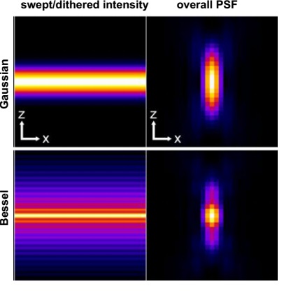
Conventional light sheets created with Gaussian beams are too thick over cellular dimensions to allow subcellular imaging. Nondiffracting beams swept across the imaging field creates a virtual light sheet of submicrometer thickness, well suited to noninvasive highspeed 4D imaging. |
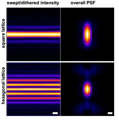
By spreading multiple ultrathin, nondiffracting light sheets of low intensity across a broad area, the intensity at any single plane is reduced, and the effect of nonlinear photodamage mechanisms is minimized.
|
Specimens are cultured or mounted on5mmdiameter cover slips. No special dye or sample treatment is required. The sample holder has inlet and output ports for perfusion of heated or cooled media.
|
|
|
|
20x increase in speed relative to spinning disc confocal microscopy |
|
The high axial and temporal resolution is achieved simultaneously - which is not possible under either the spinning disk confocal or Gaussian light-sheet paradigms. |
➸ System
3i Lattice LightSheet Microscope
Cutting edge microscopy, user friendly SlideBook™ interface
|
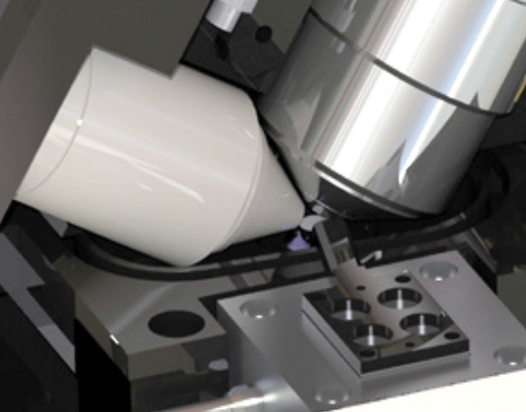
Lattice LightSheet incorporates two highly specialized objectives at right angles in an aqueous interface above the specimen. The 25x/1.1NA imaging objective is a water immersion lens with correction collar and a long2mmworking distance. A custom-designed 0.71 NA long working distance illumination objective was constructed to fit immediately adjacent to the imaging objective and perfectly illuminate the imaging field for maximum signal and resolution. A third objective is located below the specimen chamber and serves as eyepieces for sample location. |
Illumination is highly specialized for optimal power delivery in an exceptionally thin light sheet. A high-speed spatial light modulator (SLM) in combination with an annular mask allows spatially confined optical lattices to be projected onto the sample. A galvo mirror controls lattice movement, either dithering to form a uniform sheet or discretely stepping for super resolution structured illumination microscopy (SIM). |
|
|
A high-speed high-resolution sCMOS camera is used to capture image data. SlideBook™6 software controls all aspects of the system allowing for intricate high-speed synchronization of laser firing, SLM pattern display, galvo movements and camera readout while providing the ability to de-skew raw data, deconvolve and view 3D renderings of the sample. The specimen chamber and system are environmentally controlled to enable long-term living cell experiments. Access ports allow perfusion of stimulation agents, with delivery schedule digitally controlled by SlideBook™6 and synchronized with imaging. |
||
|
|
|
➸ Specifications
Technical Specifications
|
Component |
3i Lattice LightSheet |
|
Illumination Optics |
Custom lens 25x/0.71 |
|
Illumination |
- Transmission LED and epifluorescence LED for sample positioning - 405nm, 445nm, 488nm, 514nm, 561nm, 640nm |
|
Detection Modules |
Nikon 25x/1.1, WD2mm(coupled with500mmfocal length tube lens to give 62.5x) |
|
Sample Chamber |
5mm coverslip |
|
Software |
SlideBook™ |
|
Incubation |
- Perfusion specimen chamber with heating and CO2 control - Soluble factor perfusion port with Slidebook programmable scheduling |
|
Acquisition Speed |
1,000 frames/sec, 100 x 20 um in xy field of view, 3D 18,000 frames/sec, 53 x 53 um in xyfield of view, 2D |
|
SpectralRangeofDetection |
400 - 740nm |
|
Detection Zoom |
0 - 2.5x, continuous |
|
XYZ Field of View (um) |
100um x 70 um x 100um |
|
XYZ Resolution (nm) with 488nm excitation |
150 x 230 x 280 (SIM) 230 x 230 x 370 (Dither) |
|
Light Sheet Thickness (nm) |
approx 400nm |
|
Detection Module |
Hamamatsu ORCA-Flash 4.0 v2 sCMOS, can be connected to dual camera port via C-mount |
|
Pixel size Max. pixel format Bit depth Max. frame rate |
6.5um 2048 x 2048 (4.2 Mpixel) 16 bit 100fps at full frame |
➸ About 3i
INTELLIGENT IMAGING INNOVATIONS
New Innovations Address Technical Limitations
|
|
Our collective aim is to provide advanced multidimensional microscopy platforms that are intuitive to use, modular in design, and meet the evolving needs of investigators in the biological research community.
3i’s headquarters inDenver,Colorado(USA) is a repurposed taxi repair garage modernized using 100% reclaimed materials. The building features – among other recycled and reused items – hockey glass, reclaimed pallet racks, rail boxcar floorboards, and a ‘cellular’ wall system made from 20,000 recycled PET plastic water bottles.
3i’s European headquarters are located inGöttingen,Germany, home of the renown Georg-August-Universität Göttingen. Additionally, 3i has a wide global presence with application and research scientists throughout theUnited States(San Diego,Los Angeles,Baltimore) and Europe (London,Paris). 3i has partnered with numerous dealers located around the world includingChina,Japan,India,Taiwan, andIsrael. |
|
Intelligent Imaging Innovations (3i) designs and manufactures cutting edge live cell and intravital microscopy imaging platforms driven by our SlideBook™software. 3i was established in 1995 by a group of scientists whose wide range of research activities includes cell biology, immunology, neuroscience and computer science. |