

BUILT BY SCIENTISTS FOR SCIENTISTS. Intelligent Imaging Innovations (3i) designs and manufactures cutting edge live cell and intravital microscopy imaging platforms driven by 64-bit SlideBook software. 3i was established in 1995 by a group of scientists whose wide range of research activities includes cell biology, immunology, neuroscience and computer science. Our collective aim is to provide advanced multi-dimensional microscopy platforms that are intuitive to use, modular in design, and meet the evolving needs of investigators in the biological research community.
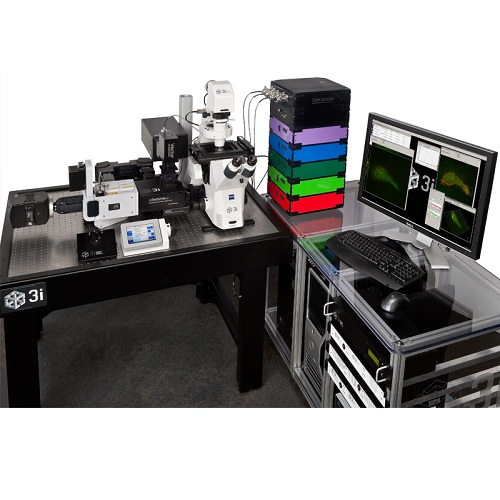
The Marianas platform incorporates advanced optics, cameras, computers and proprietary electronics to achieve unparalleled speed, precision and flexibility in live cell image acquisition. Innovative SlideBook software works seamlessly withinMarianasfor powerful and intuitive image acquisition and analysis.
An ever-growing lineup of 3i technologies and our team of experienced scientists and programmers ensure thatMarianasis always a step above the rest.
Advanced Digital Microscopy Systems
|
|
|
|
|
|
VIVO 2-Photon |
MARIANAS |
VIVO |
EVEREST |
|
|
Fixed-stage upright microscope Platform designed for highspeed multiphoton imaging of live animals and tissue |
Inverted microscope platform designed for both live and fixed cell applications |
Fixed-stage upright microscope platform ideal for intravital imaging and electrophysiology |
Upright microscope platform optimized for convenient fixed specimen work |
|
|
METHODS |
||||
|
Confocal Imaging Fluorescence Lifetime Imaging Photomanipulation/FRAP Photoablation |
Ratiometric Imaging FRET TIRF 7D Imaging |
Fluorescence Correlation Spectroscopy Optogenetics High Content Screening
|
Photoactivation Photoswitching Photostimulation |
|
|
SOFTWARE |
|
|
|
|
|
|
SlideBook software addresses the entire experimental process in biomedical imaging. From hardware device control in acquisition to mid-experiment data analysis to offline image processing and analysis, SlideBook allows users to focus on scientific investigation rather than instrument functionality.
SlideBook 5 comes standard with a large number of acquisition drivers to control over 100 devices from dozens of companies. Data can be acquired in 3D, over time, multiple color channels and as fluorescence lifetimes. Also standard are a wide variety of analytical tools for image processing, mathematical operations and a full suite of statistics functions.
SlideBook 5.5 adds numerous enhancements: • LaserStream multichannel streaming capture • Extended interoperability with MATLAB ®3D Deconvolution • 6D Multicapture allows variation in capture parameters at eachX/Y position • Improved saving of multi-gigabyte datasets
|
|||
|
Modules available for advanced applications: |
||||
|
AutoQuant Blind Deconvolution Ratio Imaging FRET Rapid 4D Streaming
|
Synchronization of TTL Devices CellNet™Pattern Recognition Photomanipulation/FRAP Imaging Stereology Super-Resolution Particle Tracking |
|||


|
|
||||||||||||||||
|
|
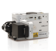
CSU-X1 • Best combination of speed and confocality for fast 3D imaging • Optimized for high NA objectives • Minimal photodamage for living cells |
|
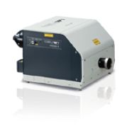
CSU-W1 • Larger field of view accommodates sCMOS cameras • Additional disk available optimized for lower NA objectives • Integrated disk movement for single camera confocal and widefield imaging |
|||||||||||||
|
|
|
|
|
|||||||||||||
|
CSU-X1 CSU-W1 |
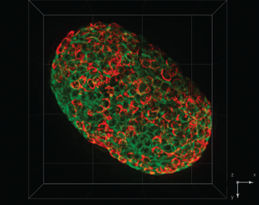
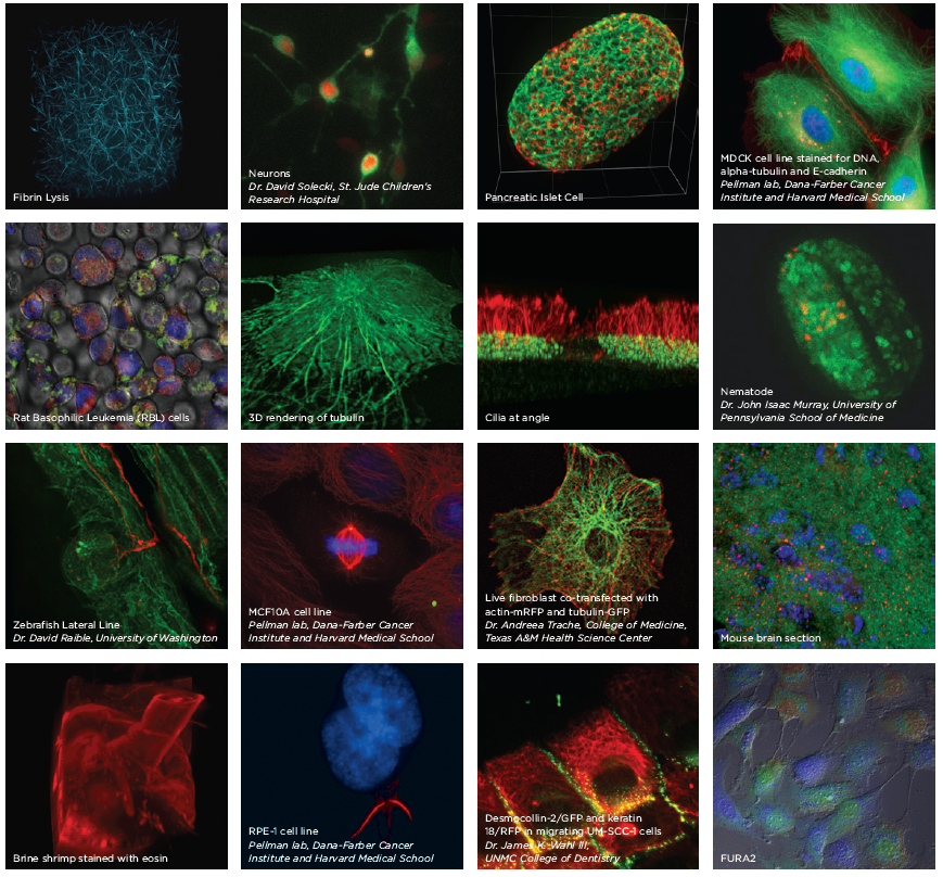
Pancreatic Islet Cell |
|||||||||||||||
|
Pinhole Diameter |
50µm disk |
25µmdisk and 50µmdisk |
||||||||||||||
|
Number of Disks |
One |
One or two with motorized switching |
||||||||||||||
|
Disk Bypass |
With 3i Bypass™ |
Standard |
||||||||||||||
|
Acquisition Speed |
2000 FPS |
200 FPS |
||||||||||||||
|
Field of View |
10mm x7mm |
17mm x16mm |
||||||||||||||
|
Near IR Excitation |
Up to 640nm |
Up to 785nm |
||||||||||||||
|
|
|
|
|
|||||||||||||
|
|
|
|
|
|||||||||||||
|
|
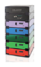
LaserStack Modular Laser Light Source • Solid state laser delivery with microsecond timing • Modular form factor allows for expansion to additional wavelengths • 405, 445, 473, 488, 514, 532, 561, 594, 640 and 660nm lines available |
|
FiberSwitcher • Sub-millisecond switch times between SDC, TIRF, FRAP andFLIM • 4 fiber-coupled outputs |
|||||||||||||
|
|
|
|
|
|||||||||||||
|
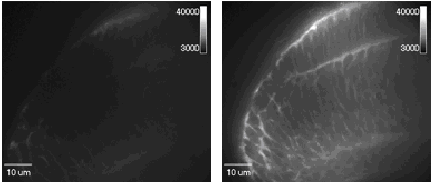
GFP mouse embryo, 100μm imaging depth, 40x objective without SAC(left), with SAC(right) |
|
mSwitcher |
||||||||||||||
|
Optical Path Selector • Millisecond switching between up to 3 devices and the microscope • 1x or 1.2x magnification(s)
|
||||||||||||||||
|
|
mSAC |
|||||||||||||||
|
Spherical Aberration Correction • Corrects for sample and preparation-induced spherical aberration • Improves geometric fidelity, resolution at depth and signal-to-noise • High speed on the fly correction as 3D data is collected
|
||||||||||||||||
|
|
Vector Modular High Speed X/Y Scanner • Accepts lasers from UV to IR • FRAP, photoactivation, photoablation, uncaging • Multiphoton beam scanning |
|
GFP in trout cells, bleached with a 488nm laser |
|||||||||||||
|
|
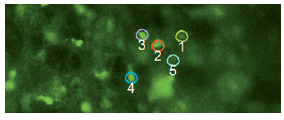
Phasor Digital Holography System • Simultaneous stimulation of non-adjacent regions in x, y and z • Optogenetic and channelrhodopsin studies |
|
preBotC network with neurons selected for simultaneous photo- stimulation via Phasor to elicit a network burst. |
|||||||||||||
|
|
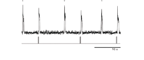
Ablate! Laser Ablation System • Attenuatable 532nm pulse laser • Localized damage tointracellular structures & targeted wound control • 70 µJ peak energy, 100Hz peak rate |
|
Bottom trace represents the laser pulse and the top trace is a recording of the hypoglossal nerve (the output of the preBotC network). Arrows note endogenous bursts; evoked bursts indicated by the laser pulse. Image and data courtesy Dr. Jason Worrell,UCLA |
|||||||||||||
|
|
|
|
||||||||||||||
|
|
Bifurcator Optical Splitter and Combiner • Creates a collimated beam space for optical accessories • Ideal for emission filter wheels • 1x or 1.2x magnification(s) |
|
Optimizer Perfect Image Relay • Optimal positioning of emission filter wheels in a collimated infinity space • 1x or 1.2x magnification |
|
mFilter Emission Filter Switcher • Millisecond switching among four positions • User-exchangeable 25mm filters • Perfect image relay |
|||||||||||
|
|
|
|
||||||||||||||
|
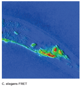
C.elegans FRET |
TIRF Total Internal Reflection Fluorescence • Manual or motorized TIRF illuminators and a selection of high NA (>1.40) objectives • Millisecond wavelength selection via LaserStack FLIM Fluorescence Lifetime Imaging • Widefield frequency-domain detection • High frequency modulated laser light source and phase-shifted intensifiedCCD detector • Combinable with widefield fluorescence, TIRF, FRAP and SDC • Sub-second acquisition FRET Förster energy resonance transfer • Supports dual camera capture and image splitting devices for real time analysis • Graphs displayed and updated during capture • Fluorescence anisotropy measurement available
|
|||||||||||||||
|
|
||||||||||||||||