


01 ABOUT
LOOK INSIDE LIVING CELLS
Every Cell is unique and has its own complex structure.
We have developed a disruptive technology which, for the first time ever, allows users to explore the inside of a living cell in 3D without the need for any labeling or other invasive methods.
The 3D Cell Explorer is a high speed, high resolution and non-invasive tool that can look deep inside biological systems. This allows us to record stunning 3D images of entire cells in just seconds and with a higher resolution than any conventional microscope on the market.
With the 3D Cell Explorer, researchers, students and medical doctors can directly experience what happens inside the living cell – in real time!

02 FEATURES
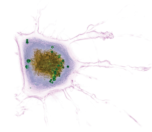
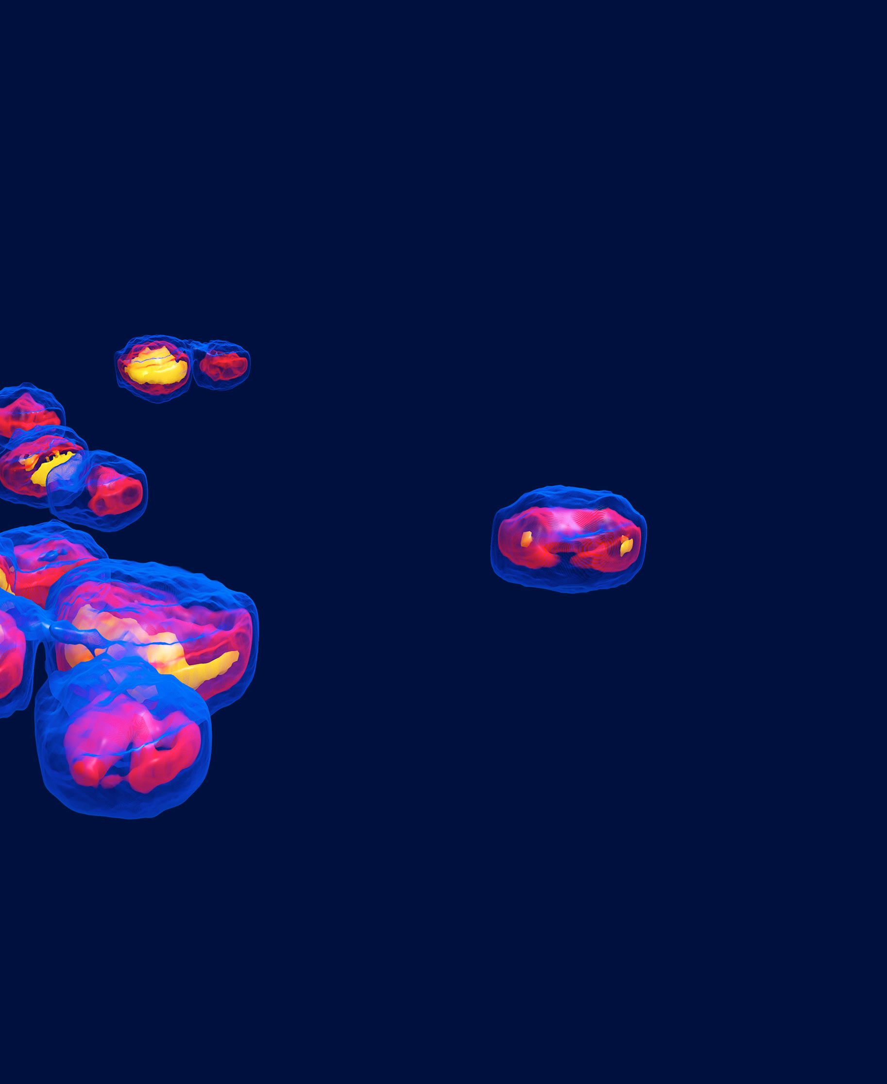
Living fibroblast reticular cell 10 μm
|
USER-FRIENDLY ACQUISITION |
NON-INVASIVE |
|
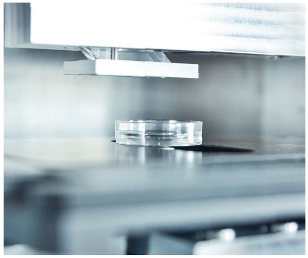
It takes just moments to get up and running with the 3D Cell Explorer: switch on the microscope, position your sample and start the acquisition with our software. Within seconds, a full 3D image of your cell will be loaded to your screen and you can start exploring.
|
Our 3D Cell Explorer delivers true live cell tomography and uses completely harmless laser light for its 3D scans of your cells. This lets you look inside a living cell without the use of labeling or markers or any other invasive methods that may potentially modify, damage or even kill the cell.
|
|
SELF-ADJUSTING |
THE FIRST UNIBODY MICROSCOPE |
|
With the 3D Cell Explorer, there is no need for sample preparation or manual calibration. After positioning your sample, the microscope will instantly self-adjust so you can be sure you are getting the best possible images of your cells.
|
The 3D Cell Explorer is the result of years of hard work and innovation and an obsession to detail. Starting from a single piece of solid aluminium, our microscope´s structure is milled down to include even the finest parts we need.
|
HOW CAN OUR MICROSCOPE BE SO AFFORDABLE?
Our goal is to give every biologist, physician, scholar and student in labs, schools and universities all over the world the chance to explore and interact directly with living cells. Thanks to a few ground-breaking innovations, which simplify but also improve our technology, we are now capable of off ering our 3D Cell Explorer at a surprisingly competitive price.
|
Marker-free imaging |
Simplified structural design |
|
Our imaging approach doesn’t rely on the weak signals encountered in traditional fl uorescence microscopy. This means we can use a commercial, industry-standard camera instead of high effi ciency scientifi c devices, even obtaining better results.
|
With our self-adjusting optics (patent
|
|
Numerical correction |
|
|
Using our patented processing technique based on complex deconvolution, we can correct for many imaging errors that otherwise require extremely expensive optical components and ultra-precise alignment.
|
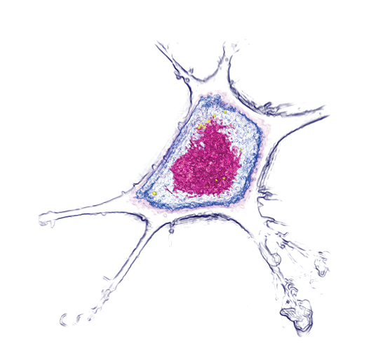
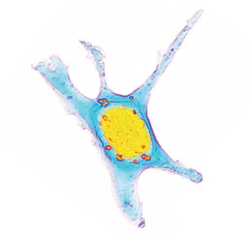
Living fibroblast reticular cell 10 μm
03 TECHNOLOGY
LIVE-CELL TOMOGRAPHY
The Cell Explorer’s technology is unique worldwide and is based on a fundamen tal patent (US & EU WO 2011/121523). The combination of holography and rotational scanning allows determation of how light propagates through the cell. By this means, we can measure the cell’s physical properties, through its refractive index. The result is quantitative cell tomography, in vitro without any invasion or sample preparation.
TECHNICAL SPECIFICATIONS
Resolution: Δx,y = 200nm; Δz = 500nm
Field of view: ~ 80µm
Depth of field: ~ 30µm
Tomography frame rate: 0.6fps 3D image rate, with full self-adjustement
Microscope objective:
Air with 60x Magnification
Low power laser (λ = 520nm, sample exposure <20nW/µm 2)

04 SOFTWARE
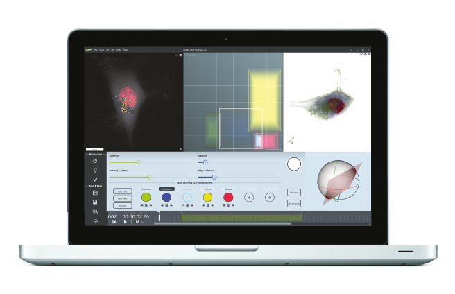
The software STEVE and its user interface.
MEET STEVE
STEVE is the Cell Explorer’s software counterpart and all you need to dive into the microcosm of living cells. Harnessing your computer’s graphic processor, we designed STEVE to run smoothly, even during acquisition. Use STEVE’s intuitive interface allows you to control the microscope, explore your data using interactive digital staining and even perform quantitative analyses on your measurements. If you are happy with your results, you can easily share them online or directly have them 3D-printed.
SOFTWARE FEATURES
• GPU-accelerated 3D processing and display
• Intuitive graphical user interface
• Quantitative staining based on physical markers (refractive index)
• Versatile statistical analysis tools
05 APPLICATIONS
A TOOL FOR DISCOVERY
The combination of holography and rotational scanning makes Nanolive’s a revolutionary technology that opens up new doors for observing seeing living cells in nanometric detail. It allows the measurement of cellular processes and kinetics in real-time enabling multi parameter analysies at single-cell and sub-cellular scales.
The 3D Cell Explorer is a tool of discovery and we are just at the beginning of exploring all the potential fields of application. There are no boundaries.
POTENTIAL FIELDS OF DISCOVERY
· Cell division
· Cell morphology monitoring
· Cell differentiation (200+ types)
· Cell-cell interaction
· Intracellular trafficking
· Cellular remodeling processes
· Cell death (apoptosis or necrosis)
· Drug monitoring